
Fig. 8
- ID
- ZDB-FIG-150326-49
- Publication
- Skaggs et al., 2014 - Excitotoxic brain injury in adult zebrafish stimulates neurogenesis and long-distance neuronal integration
- Other Figures
- All Figure Page
- Back to All Figure Page
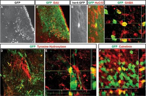
Neurons generated following QA-induced lesions of adult zebrafish telencephalon adopt multiple phenotypes. (A, A2) In Tg(her4:creERT2;β-actin2:LCLG) (her4:GFP) fish at 28 days post-lesioning, cells birthdated with EdU (A; red in A2) at 2 days after QA and tamoxifen injection have settled in the periventricular region and parenchyma of the damaged hemisphere where many coexpress GFP (green in A2). (B–B′) Higher magnification view of a GFP/HuC/D double-labeled cell with neuronal morphology in the parenchyma. (C) Neurons labeled for the inhibitory neuronal marker GABA include cells that coexpress GFP that comingle closely with other GABA-expressing cells. (D,E) Neurons double-labeled (yellow arrows) for GFP (green) and tyrosine hydroxylase (TH) comingle among the distinctive TH positive cluster of cells (red) in the ipsilesional hemisphere. (F) Within a calretinin-expressing group of neurons, some cells coexpressed calretinin (red) and GFP (green) whereas neighboring cells showed immunoreactivity for one of the two markers. Scale bar A, B, D = 100 µm. Scale bar C, E, F = 50 µm. |