
FIGURE
Fig. 8
- ID
- ZDB-FIG-150325-42
- Publication
- Kenyon et al., 2015 - Zebrafish Rab5 Proteins and a role for Rab5ab in nodal signalling
- Other Figures
- All Figure Page
- Back to All Figure Page
Fig. 8
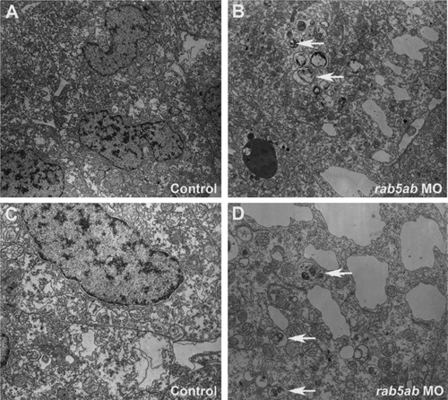
Activity of rab5ab in endocytosis. Transverse sections of cells of the leading edge of the enveloping layer from a 3 ng control-injected embryo 80% epiboly (A) and (C), a 3 ng rab5ab MO injected embryo (fixed at 40% epiboly but when control embryos were at 80% (B) and (D). White arrows show large secondary lysosomes with membranous contents. (A) and (B) are at 10,000×; and (C) and (D) are at 18,750×. |
Expression Data
Expression Detail
Antibody Labeling
Phenotype Data
| Fish: | |
|---|---|
| Knockdown Reagent: | |
| Observed In: | |
| Stage: | 30%-epiboly |
Phenotype Detail
Acknowledgments
This image is the copyrighted work of the attributed author or publisher, and
ZFIN has permission only to display this image to its users.
Additional permissions should be obtained from the applicable author or publisher of the image.
Reprinted from Developmental Biology, 397(2), Kenyon, E.J., Campos, I., Bull, J.C., Williams, P.H., Stemple, D.L., Clark, M.D., Zebrafish Rab5 Proteins and a role for Rab5ab in nodal signalling, 212-24, Copyright (2015) with permission from Elsevier. Full text @ Dev. Biol.