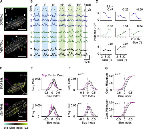
RGC Inputs to Tectal Neuropil Carry Small- and Large-Size-Selective Signals (A) Two optical sections from tectal neuropil with GCaMP6m expressed in RGC fibers. Imaging depth is 18 µm and 51 µm from the dorsal crest. Dashed lines indicate superficial, central, and deep layers. Colored boxes indicate eight representative ROIs. Arrowheads mark skin fluorescence. M, medial direction; C, caudal direction. (B) Normalized fluorescence signals from ROIs in response to moving targets and full-field flash. Object size is indicated above the stimulus trace. Numbers refer to ROIs indicated in (A). Note the whole-field flash OFF response in traces 6 and 8. (C) Size-tuning curves calculated from signal variance in ROIs indicated in (A). Variance was measured in intervals indicated by the blue (small-stimulus size) and green (large-stimulus size) vertical boxes in (B). Size index (S.I.) was calculated as S.I. = (Varlarge Varsmall) / (Varlarge + Varsmall). (D) Size selectivity maps for optical sections shown in (A) with corresponding color scale. (E) Normalized frequency distributions of size indices determined from single-pixel fluorescence signals, sorted for the superficial, center, and deep layers of the neuropil and displayed separately for the dorsal and ventral imaging plane. Data are from (D). (F) Average frequency distributions of size indices from 11 fish, for dorsal and ventral imaging planes. (G) Cumulative histograms of size indices. The same data as in (F) are shown. Scale bars, 50 µm (A and D). See also Figure S1.
|
