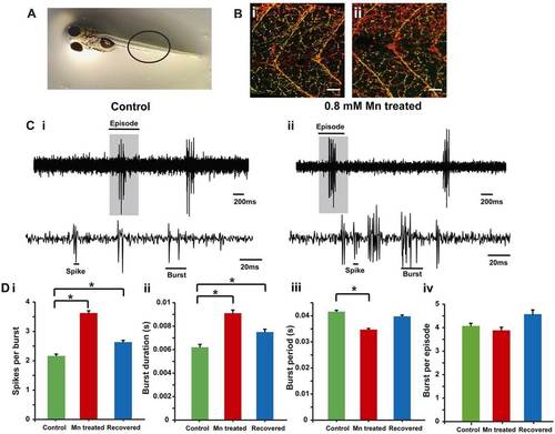
Morphology and function of motor neurons. (A) Zebrafish larvae, the region where ZNP-1 antibody and α-bungarotoxin staining was visualized (black circle) is highlighted. (B) A single optical slice showing ZNP-1 staining (red) and α-bungarotoxin staining (green, colocalized as yellow) of motor neuron innervation of axial muscles in (i) control and (ii) Mn-treated (48-hour exposure) larvae. The staining indicates similar motor neuronal axon morphology between the two groups. Scale bars: 30 µm. (C) Extracellular recordings of fictive swim patterns on motor neurons highlight that episodes comprise bursts and spikes in (i) control and (ii) Mn-treated larvae. The top traces show episodes of swim motor patterns. The episode highlighted by a gray box is shown expanded in the bottom traces. (D) Bar plots for (i) the number of spikes per burst, (ii) the burst duration, (iii) the burst period and (iv) the number of bursts per episode in control (green), Mn-treated (red) and recovered (blue) larvae. Data represent mean±s.e.m. *Values that are significantly different from control at an α level of 0.05 as estimated by using the Kruskal–Wallis test.
|
