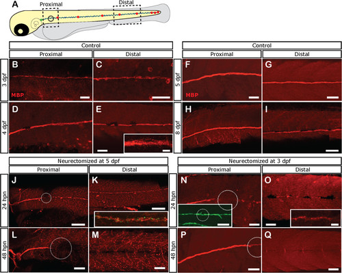
Loss of Schwann cell differentiation markers after denervation. Tg(foxD3:GFP) larvae were left untreated or neurectomized and then fixed at different times and processed for immunostaining with anti-Myelin Basic Protein (MBP; labeled in red). (A) The expression of MBP was evaluated in a proximal and distal area with respect to the point of neurectomy (black circle). (B-I) In control larvae. MBP is expressed at proximal and distal levels at 3 dpf and increases as the larva ages. (J-M) Expression of MBP becomes absent after neurectomy in denervated Schwann cells. Five-day-old tg(NeuroD:GFP) larvae were neurectomized and were fixed and processed for immunostaining with anti-MBP 1 (J, K) or 2 days (L, M) after neurectomy. Nerve degeneration (green label in inset in K) is followed by fragmentation of MBP expression in Schwann cells distal to the injury point (white dotted circle) at 24 hpn (J, K, inset in K shows both channels). At 48 hpn (L, M), MBP expression has disappeared distal to the injury site. (N-Q) Neurectomy at earlier stages, when MBP becomes expressed (at 3 pdf), showed the same results. In this case, Tg(foxD3:GFP) larvae were used to follow Schwann cells; note that despite fragmentation and loss of the MBP label, Schwann cells still remain viable as revealed by GFP expression. Scale bars: J, K, inset in N: 100 µm; B-I, L-Q, inset in E, inset in O: 50 µm.
|
