
Fig. 1
- ID
- ZDB-FIG-150115-29
- Publication
- Fukuhara et al., 2014 - Visualizing the cell-cycle progression of endothelial cells in zebrafish
- Other Figures
- All Figure Page
- Back to All Figure Page
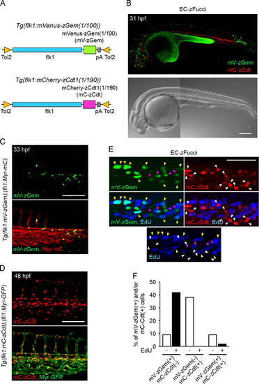
Development and characterization of transgenic zebrafish lines expressing zFucci cell-cycle biosensors in endothelial cells (ECs). (A) Schematic representation of the plasmids used for delivering transgenes expressing mVenus-zGem (1/100) (mV-zGem) and mCherry-zCdt1(1/190) (mC-zCdt) under control of the flk1 promoter. (B) Upper, fluorescence image (mV-zGem [green] and mC-zCdt1 [red]) of an EC-zFucci, Tg(flk1:mV-zGem);(flk1:mC-Cdt) embryo, at 31 hpf; lower, bright field images. (C) 3D-rendered confocal stack fluorescence images of the caudal regions of the Tg(flk1:mV-zGem);(fli1:Myr-mC) embryo at 33 hpf. Upper, mVenus image; lower, the merged image of mVenus (green) and mCherry (red). All of the confocal fluorescence images are lateral views and displayed as anterior to the left, unless otherwise described, in the following images. (D) 3D-rendered confocal images of the caudal regions of the Tg(flk1:mC-Cdt);(fli1:Myr-GFP) embryo at 48 hpf. Upper, mCherry image; lower, the merged image of mVenus (green) and mCherry (red). (E) Images of the EC-zFucci embryo at 29.5 hpf treated with EdU for 1 h. mVenus images (green), mCherry images (red) and EdU images visualized by Alexa 647-azide (blue) of the trunk vessel are shown as indicated at the lower left corner of the image. Yellow, pink and white arrowheads indicate mV-zGem/EdU double-positive cells, mV-zGem-positive/EdU-negative cells and mC-zCdt-positive/EdU-negative cells, respectively. (F) Percentages of mV-zGem-positive, mC-zCdt-positive and mV-zGem/mC-zCdt double positive cells labeled without () or with (+) EdU as observed in E were quantified (n=55). Scale bars, 200 μm (B) and 100 μm (C–E). |
| Genes: | |
|---|---|
| Fish: | |
| Anatomical Terms: | |
| Stage Range: | Prim-5 to Long-pec |
Reprinted from Developmental Biology, 393(1), Fukuhara, S., Zhang, J., Yuge, S., Ando, K., Wakayama, Y., Sakaue-Sawano, A., Miyawaki, A., Mochizuki, N., Visualizing the cell-cycle progression of endothelial cells in zebrafish, 10-23, Copyright (2014) with permission from Elsevier. Full text @ Dev. Biol.