
FIGURE
Fig. 2
- ID
- ZDB-FIG-141006-15
- Publication
- Moshal et al., 2011 - Discriminating Different Cancer Cells Using a Zebrafish in Vivo Assay
- Other Figures
- All Figure Page
- Back to All Figure Page
Fig. 2
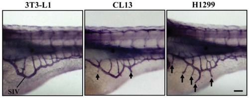
Xenotransplantation-induced neovascularization in the zebrafish embryos. Lateral views of whole-mount alkaline phosphatase stains of zebrafish embryos at 3 dpf were used to evaluate 3T3-L1 control, CL13 and H1299 cancer cell lines for angiogenic potential by measuring newly formed ectopic vessels sprouting from the SIV plexus in the zebrafish/tumor xenograft assay. They show negligible, moderate, and robust angiogenic responses, respectively. Scale bar = 100 μm. |
Expression Data
Expression Detail
Antibody Labeling
Phenotype Data
Phenotype Detail
Acknowledgments
This image is the copyrighted work of the attributed author or publisher, and
ZFIN has permission only to display this image to its users.
Additional permissions should be obtained from the applicable author or publisher of the image.
Full text @ Cancers