
FIGURE
Fig. S1
Fig. S1
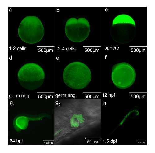
Staining pattern of DCFH-DA at early stages. (a-h) The staining pattern of DCFH-DA at different stages. g2 is the high magnification of boxed place in g1 from ventral-dorsal angle, which shows the opening of the pronephric ducts through cell apoptosis. |
Expression Data
Expression Detail
Antibody Labeling
Phenotype Data
Phenotype Detail
Acknowledgments
This image is the copyrighted work of the attributed author or publisher, and
ZFIN has permission only to display this image to its users.
Additional permissions should be obtained from the applicable author or publisher of the image.
Full text @ Sci. Rep.