
Fig. 7
- ID
- ZDB-FIG-140917-45
- Publication
- Carr et al., 2014 - Characterization of the Zebrafish Homolog of Zipper Interacting Protein Kinase
- Other Figures
- All Figure Page
- Back to All Figure Page
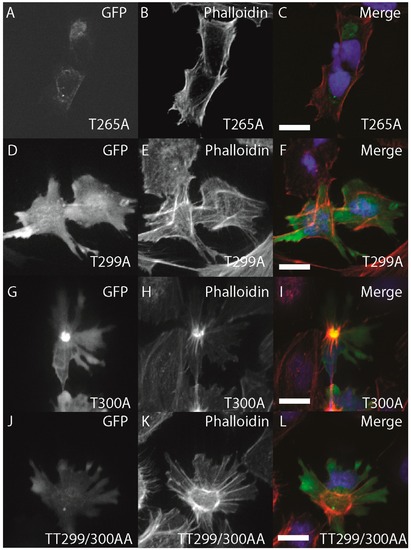
Structure-function of zebrafish ZIPK. HeLa cells were transfected with either GFP-ZIPK constructs generated by site-directed mutagenesis mutating a proposed activating phosphorylation site T265 or phosphorylation sites involved in controlling subcellular localization in the human paralog T299 and T300. All cells were fixed and stained with DAPI and Alexa 568-phalloidin. (A, D, G, J) show phalloidin staining; (B, E, H, K) show GFP; while color images (C, F, I, L) are a merge of DAPI (blue), GFP (green) and phalloidin (red). Each experiment was performed at least five times with a minimum of 25 cells assayed per experiment. White bar indicates 20 μm. |