
Fig. 5
- ID
- ZDB-FIG-140819-6
- Publication
- Trevarrow et al., 1990 - Organization of hindbrain segments in the zebrafish embryo
- Other Figures
- All Figure Page
- Back to All Figure Page
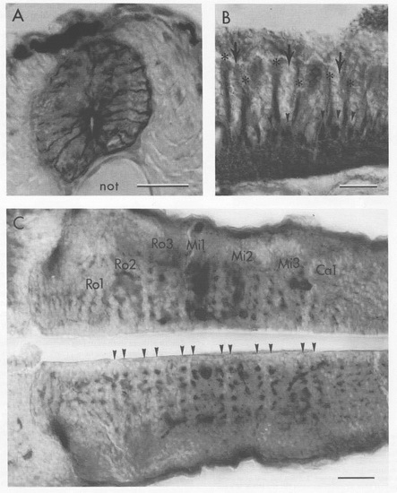
Rows of Radial Glial Fibers Separate the Segment Centers from Their Borders (A) The zrf-1 antibody labels typically shaped radial glial fibers that extend from the cells at the central canal to the pial surface in this transverse section of a 4 day zebrafish spinal cord. not: notochord. The spinal cord and hindbrain were labeled similarly by the other rf antibodies and by a mouse monoclonal antibody (from ICN Immunobiological) to GFAP (data not shown). (B) In the hindbrain, bundles of dorso-ventrally running glial fibers (asterisks) are present between the segment borders (large arrows) and centers. This sagittal section, at 48 hr, was labeled with a mixture of zrf-1, zrf-2, and zrf-4 antibodies to stain the glial fibers heavily and with zn-5 to label commissural axons (arrowheads). The axon bundles run orthogonally to the glial fiber bundles, and the two appear to be in close contact. (C) The glial bundles, cut across in this horizontal sec8tion and labeled with zrf-1 at 48 hr, are arranged in parallel transverse rows (arrowheads), two in each segment. Retrogradely labeled reticulospinal neurons are dorkly stained in the Mi1 and Mi2 segments and mark the locations of he segment centers. The hindbrain-midbrain boundary is to the left. Scale bars: 25 μm. |
| Antibodies: | |
|---|---|
| Fish: | |
| Anatomical Terms: | |
| Stage Range: | Long-pec to Day 4 |