
Fig. 6
- ID
- ZDB-FIG-140812-32
- Publication
- Kessels et al., 2014 - Proteomics analysis of the zebrafish skeletal extracellular matrix
- Other Figures
- All Figure Page
- Back to All Figure Page
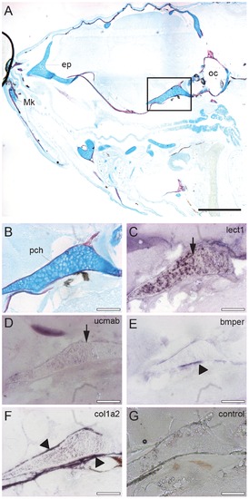
In situ mRNA hybridization of genes encoding extracellular proteins implicated in cartilage and bone formation in the head region. Sagittal sections of the head region of 28 dpf zebrafish juveniles stained by acid-free bone (red) and cartilage (blue) staining (A-B), or used for in situ hybridization with an antisense RNA probe corresponding to lect1 (C), ucmab (D), bmper (E), col1a2 (F). As a control, hybridization with the lect1 sense RNA probe is shown (G). (B-G) Magnification of boxed area in A focusing on the parachordal (pch) cartilage. Images are from consecutive sections at the same position. (C-D) Expression of lect1 and ucmab is located in chondrocytes of the parachordal (pch) cartilage (arrows). (E-F) Transcripts of bmper and col1a2 were detected in ossification sites surrounding the parachondral chondrocytes (arrow heads). Scale bars indicate 0.5 mm (black), or 100 µm (white). Abbreviations: ep, ethmoid plate; Mk, Meckel’s cartilage; oc, otic capsule; pch, parachordal. |
| Genes: | |
|---|---|
| Fish: | |
| Anatomical Term: | |
| Stage: | Days 21-29 |