
Fig. 5
- ID
- ZDB-FIG-140728-13
- Publication
- Asaoka et al., 2014 - The Hippo Pathway Controls a Switch between Retinal Progenitor Cell Proliferation and Photoreceptor Cell Differentiation in Zebrafish
- Other Figures
- All Figure Page
- Back to All Figure Page
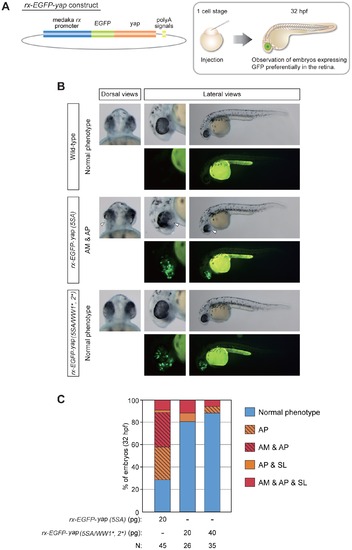
Retina-specific expression of yap (5SA) induces retinogenesis defects without affecting body axis formation. (A) Schematic illustration of the base rx-EGFP-yap construct (left panel) and the procedure for the experiment (right panel). Zebrafish embryos at the one-cell stage were injected with plasmid DNA containing the rx promoter constructs indicated in (B). (B) Representative dorsal and lateral views of the embryos in (A) that were injected with rx promoter constructs as indicated on the left side of panels. Data are presented as for Fig. 4B. White arrowheads, areas of AM plus AP. (C) Quantification of phenotypes of the embryos injected with rx promoter constructs in (A, B) as analyzed at 32 hpf. Color classification is as for Fig. 4C except that the phenotype of AM plus AP is indicated by striped red shading. Results are presented as the percentage of the total number of embryos examined (N). Note that expression of yap (5SA) variants mutated in both the WW1 and WW2 domains prevented the appearance of abnormal eye phenotypes. |