
Fig. S3
- ID
- ZDB-FIG-140626-35
- Publication
- West et al., 2014 - Unusual Fluorescent Granulomas and Myonecrosis in Danio Rerio Infected by the Microsporidian Pathogen Pseudoloma Neurophilia
- Other Figures
- All Figure Page
- Back to All Figure Page
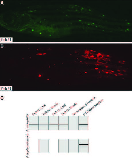
PCR analysis of microsporidia-infected D. rerio. Two fish with fluorescent granulomas were commercially assayed by PCR for microsporidial DNA from Pseudoloma neurophilia and Pleistophora hyphessobryconis (IDEXX RADIL, Columbia, MO). (A) Lck:EGFP transgenic fish with fluorescent nodules, wasting, and abnormal spine curvature imaged using FITC/GFP filter (emission spectrum 500–550 nm). (B) Identical fish imaged using TRITC filter (emission 575–625 nm). Like other animals harboring fluorescent nodules (Fig. 1), lesions coincide using either filter, chiefly in skeletal muscle of flanks. (C) PCR results visualized by capillary electrophoresis: P. neurophilia was detected in CNS and skeletal muscle of both fish (top, lanes 1–4). No template (lane 5, negative control) shows no product; positive control (lane 6) yielded band of identical size. P. hyphessobryconis, a Microsporidian known to infect muscle, was not detected in skeletal muscle of either fish (bottom, lanes 2 and 4). CNS, central nervous system; FITC, fluorescein isothiocyanate; PCR, polymerase chain reaction. |