
Fig. S2
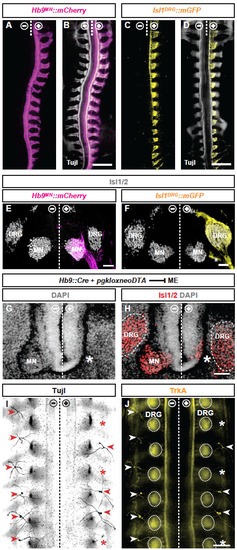
Visualizing and manipulating peripheral MEs and SAs in chick. (A) Dorsal wholemount view of MEs (magenta: Hb9MN::mCherry) in E5 chick embryo (-/+: untransfected/transfected hemitubes). (B) Overlay of MEs with pan-axon label (grey: anti- Tuj-1 immunofluorescence). (C) Dorsal wholemount view of SAs (yellow: Isl1DRG::mGFP) in E5 chick embryo. (D) Overlay of SAs with Tuj-1 immunofluorescence. (E) Transversal section of E6 chick neural tube: unilaterally Hb9MN::mCherry-labelled MEs extending from motor neurons (MNs). Overlay with anti-Isl1 immunofluorescence (grey) to label DRG neurons and MNs. (F) Transversal section of neural E6 chick tube: Isl1DRG::mGFP-labeled SAs extending from DRG neurons, in addition to a subset of non-DRG cells (presumably of neural crest origin) also labeled by the Isl1DRG enhancer (asterisk). (G,H) Transversal section of E5 chick spinal cord: severe reduction of MN numbers, including loss of ventral horn (asterisk), but not DRG neuron numbers, upon unilateral Hb9MN::Cre/PGKneolox2DTAmediated MN ablation (grey: cell nuclei labeled by DAPI). (I) Dorsal wholemount view of E7 chick embryo after unilateral Hb9MN::Cre/PGKneolox2DTA-mediated MN ablation. Innervation of dermis by dorsal cutaneous nerves (arrowheads) visualized by anti-Tuj-1 immunofluorescence (black): intermittent loss of dorsal cutaneous nerves in Hb9MN::Cre/PGKneolox2DTA-transfected (+), but not control (-) side. (J) Visualization of most DRG neurons and SAs by anti-TrkA immunofluorescence (yellow): loss of dorsal cutaneous SA projections (asterisk), but not DRGs (encircled by dotted lines). Scale bars: 300 µm in B,D,J; 50 µm in E,F,H. |