
Fig. 1
- ID
- ZDB-FIG-140522-21
- Publication
- Takayama et al., 2014 - Expression patterns of dnmt3aa, dnmt3ab, and dnmt4 during development and fin regeneration in zebrafish
- Other Figures
- All Figure Page
- Back to All Figure Page
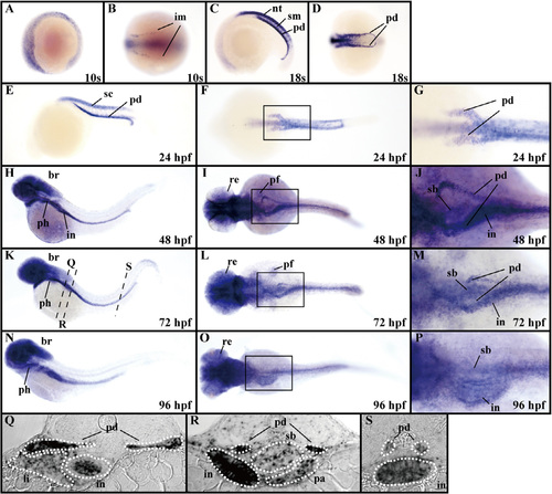
Expression patterns of dnmt3aa in zebrafish embryos and larvae. (A–P) dnmt3aa expression was examined by whole-mount in situ hybridization at the 10s and 18s stages as well as at 24, 48, 72, and 96 hpf. Lateral views, anterior to the left (A, C, E, H, K, and N). Vegetal pole views of the tail-bud region, anterior to the left (B and D). Dorsal views, anterior to the left (F, I, L, and O). The boxed areas in (F), (I), (L), and (O) are shown enlarged in (G), (J), (M), and (P), respectively. (Q–S) Transverse sections showing dnmt3aa expression. The transverse sections were cut at the levels indicated by the dashed black lines in (K). br, brain; im, intermediate mesoderm; in, intestine; li, liver; nt, neural tube; pa, pancreas; pd, pronephric duct; pf, pectoral fin buds; ph, pharyngeal arches; re, retina; sb, swim bladder; sc, spinal cord; sm, somitic mesoderm. |
| Gene: | |
|---|---|
| Fish: | |
| Anatomical Terms: | |
| Stage Range: | 10-13 somites to Day 4 |
Reprinted from Gene expression patterns : GEP, 14(2), Takayama, K., Shimoda, N., Takanaga, S., Hozumi, S., and Kikuchi, Y., Expression patterns of dnmt3aa, dnmt3ab, and dnmt4 during development and fin regeneration in zebrafish, 105-110, Copyright (2014) with permission from Elsevier. Full text @ Gene Expr. Patterns