
Fig. S3
- ID
- ZDB-FIG-140512-50
- Publication
- Dreosti et al., 2014 - Left-right asymmetry is required for the habenulae to respond to both visual and olfactory stimuli
- Other Figures
- All Figure Page
- Back to All Figure Page
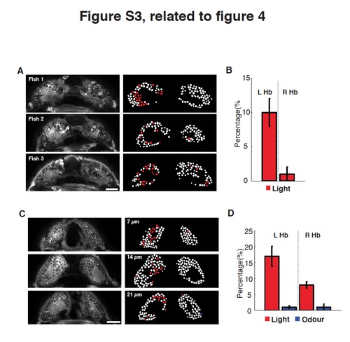
Dark-reared fish and fish with olfactory inputs removed show no change in lateralization of functional sensory responses. (A-D) Examples (A) of a two-photon images of a single planes of the dHb (at 14 μm depth), where the majority of light responses are localised, of three Tg(elavl3:GCaMP5G) 4 dpf dark- reared fish (left). Corresponding colour-coded calcium signals in response to a light stimuli (right). Light responding neurons are in red. Scale bar 20μm. (B) Bar chart showing the percentages of light responding neurons (normalized to the total number of dHb neurons) in the left and right dHb nuclei for two sampled planes (at depths of 7 and 14μm) in multiple dark-reared fish (number of fish=4). (C) Examples of two-photon images of three single planes of the dHb (7, 14 and 21 μm depth) of a Tg(elavl3:GCaMP5G) 4 dpf fish with nose pit removal at 1 dpf, before mitral cell projections reach the Hb, (left). Corresponding colour-coded calcium signals in response to non-lateralized presentation of light and odour (right). Light responding neurons are in red, while odour responses in blue. Scale bar 20μm. (D) Bar charts showing the percentages of light (red) versus odour (blue) responding neurons (normalized to the total number of dHb neurons) in the left and right dHb nuclei for two sampled planes (at 7 and 14μm, number of fish=6). Functional asymmetry of light responses is still present after olfactory pit removal. Error bars are SEM. |