
Fig. 4
- ID
- ZDB-FIG-140409-44
- Publication
- Varga et al., 2014 - Autophagy is required for zebrafish caudal fin regeneration
- Other Figures
- All Figure Page
- Back to All Figure Page
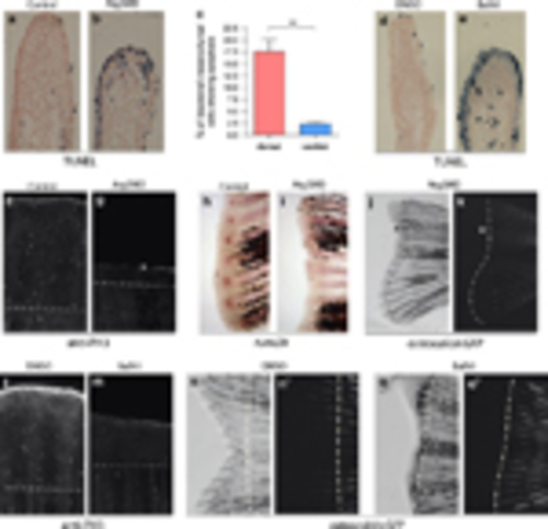
Reduced autophagic activity impairs cell survival, proliferation and differentiation during regeneration. (a–c) When dorsal 2-dpa blastema cells were injected with Atg5MO and fixed at 6hpi, TUNEL staining showed increased cell death in the blastemal mesenchyme of treated fin halves, suggesting that autophagy is required for the survival of dedifferentiating cells. (For the statistical analysis apoptotic and non-apoptotic cells were counted on 62 ventral and 49 dorsal sections, **P<0.01, paired t-test.) (d and e) Chronic treatment with bafilomycin A1 also results in reduced regeneration and increased cell death at 2dpa in the blastemal mesenchyme. (f and g) At 3dpa significant amount of mitotic cells, marked by anti-phospho-histone 3 staining can be detected in a control blastema, whereas the number of proliferating cells is significantly reduced in Atg5MO-injected blastemas. (h and i) Six hours post injection 2-dpa caudal fins were fixed and stained for runx2b expression. Whereas the uninjected ventral half shows runx2b staining in the blastema at the tip of the individual fin rays (h), in injected dorsal blastema no staining could be observed (i), suggesting that the survival of pre-osteoblasts was impaired upon morpholino treatment. (j and k) An amputated adult fish shows no osteocalcin:GFP expression at 6dpa in the Atg5MO-treated dorsal areas. White lines indicate the extent of GFP expression in the bright field (left) and the corresponding fluorescent image (right). The asterisk indicates parts of the blastema tissue that fail to express osteocalcin. (l and m) Chronic treatment with bafilomycin A1 decreases the number of mitotic cells at 3dpa. (n–o2) Long-term impairment of autophagic activity with bafilomycin A1 suppresses (re)differentiation as shown by the osteocalcin:GFP reporter line in 6dpa regenerates. Unlike in controls (n2), in treated fish no new osteocalcin-positive cells could be observed at 6dpa in the fin (o2) (Dashed lines denote the plane of amputation.) |