
Fig. S2
- ID
- ZDB-FIG-140325-39
- Publication
- Priyadarshini et al., 2013 - Oxidative stress and regulation of Pink1 in zebrafish (Danio rerio)
- Other Figures
- All Figure Page
- Back to All Figure Page
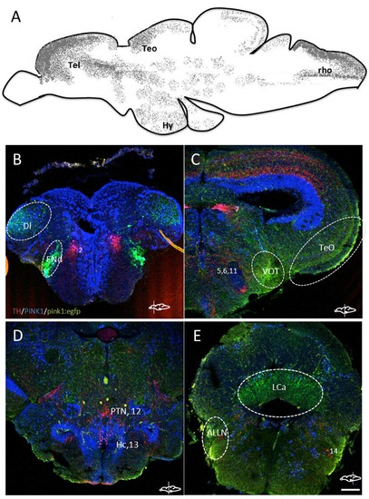
A. Schematic lateral view of pink1:egfp expressing regions in the zebrafish brain compiled from immunohistochemistry of larval and adult brain sections. The regions are marked as: Tel – telencephalon, Teo – anterior region of the optic tectum, Hy – hypothalamus, rho – rhombencephalon. B-E. Cryosections of different regions of the adult zebrafish brain with immunoreactivity detected for TH (red), PINK1 (blue) and pink1:egfp (green). The cell populations of TH-ir are reported with numbers, and additional regions of GFP are marked with dotted lines. Di – lateral zone of the dorsal telencephalon, ENd – endopeduncular nucleus, VOT – ventrolateral optic tract, TeO – tectum opticum, PTN – posterior tuberal nucleus, Hc – caudal zone of the periventricular hypothalamus, LCa – lobus caudalis cerebelli, ALLN – anterior lateral line nerves. Scale bar represents 100 μm. |