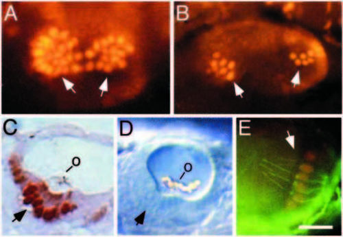
Overproduction of hair cells in dlAdx2/dx2 mutants. (A) Dorsolateral view of the otic vesicle of a 30 h dlAdx2/dx2 embryo stained with anti-Pax2 antibody. Hair cells stain intensely (arrows) and are produced in much greater numbers than normal. (B) Dorsolateral view of the otic vesicle of a 30 h dlAdx2/T(msxB)b220 trans-heterozygote stained with anti-Pax2 antibody. Stained hair cells (arrows) are produced in excess, but to a lesser degree than in dlAdx2/dx2 homozygotes. (C) Parasagittal section of a dlAdx2/dx2 embryo stained in whole mount with anti-Pax2 and anti-acetylated tubulin antibodies. Large numbers of hair cells are labeled (arrow), but few support cells are evident. An otolith (o) is attached to the hair cell ciliary bundles. (D). Lateral view of the otic vesicle of a live dlAdx2/dx2 embryo as seen under DIC optics. Numerous hair cells are evident (arrow) and a large malformed otolith (o) is distributed across the tips of the hair cell ciliary bundles. (E) Lateral view of the posterior crista of a 60 h dlAdx2/dx2 embryo stained in with anti-Pax2 (red) and anti-acetylated tubulin (green) antibodies. Hair cells show nuclear Pax2 staining and are produced in greater than normal numbers. Anterior is to the right and dorsal is to the top. Scale bar, 15 μm (E), 20 μm (A-C), or 25 μm (D).
|
