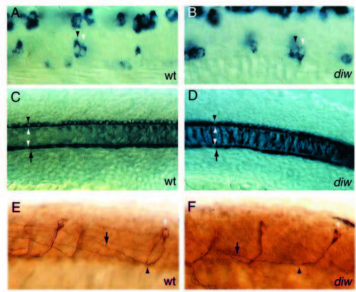
Motoneuron specification, notochord and floorplate development and spinal axonal projections are not affected in diwanka mutant embryos. (A,B) islet-1-positive cells in the ventral spinal cord consistent with the position of RoP neurons (black arrowhead) and MiP neurons (white arrowhead) are present in wild-type and mutant embryos. CaP, MiP and RoP motoneurons were identified by their dorsoventral and rostrocaudal position within the spinal cord and their soma size (see Table 1 for details). (C,D) type II collagen expression in the notochord (delineated by white arrowheads), in the floorplate (between black arrowhead and upper white arrowhead) and hypochord (between arrow and lower white arrowhead) is indistinguishable between wildtype and mutant embryos, suggesting that these cell types are not affected by mutations in the diwanka gene. (E,F) In wild-type and mutant embryos 3A10-positive commissural spinal interneurons (white arrowhead) extend their axons ventrally to the midline (black arrowhead), cross the midline and ascend on the contralateral side (arrow). Multiple focal planes were compiled to follow the entire axonal projections.
|
