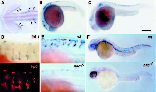
Expression of the zebrafish Mitf-related gene during development. Wholemount in situ hybridization was performed with an antisense probe to 3A.1. (A) Expression (arrowheads) in the caudal margin of the eye and in head neural crest of an 18-somite (18 hpf) embryo. Expression is also detectable in a few cells in the trunk neural crest at this stage (not shown). 3A.1 expression expands in the eye and progresses in the head and trunk in a rostral to caudal manner as seen in 21 hpf (B) and 23 hpf (C) embryos. Migratory cells can clearly be seen by the latter timepoint. (D) 3A.1 (blue, top panel) and the melanoblast marker trp2 (red, bottom panel) are coexpressed in migrating melanoblasts of 24 hpf embryos. (E) Expression of 3A.1 is reduced in nacre mutants. The top panel shows a closeup of the 23 hpf embryo from (C). 3A.1-expressing cells can be seen on migratory paths at each somite level. Expression is much reduced and few cells have migrated away from the neural tube in a nacre mutant embryo at the same stage (bottom panel). (F) Reduced 3A.1 expression is still clearly evident at 30 hpf in a comparison of albino (top) and nacre (bottom) embryos. Scale bar: A, 100 μm; B,C, 200 μm; D,E, 50 μm; F, 250 μm.
|

