|
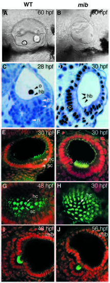
Wild-type and mib ears compared. (A,B) Lateral views of live embryos (Nomarski optics) at 60 hpf. Note that the ears are smaller and deformed in mib and that the otoliths are drastically reduced, reflecting a deficit in the sensory patches. (C-D) Semi-thin Araldite sections stained with toluidine blue, showing the altered pattern of cell differentiation. (E-H) Confocal images of whole-mount specimens stained with fluorescent phalloidin (green) to reveal actin-rich hair bundles and with 7AAD (red nuclear staining) as a counterstain. (E,F) Optical sections passing transversely through sensory patches; (G,H) En-face views of sensory patches. Supporting cells are missing in mib, while hair cells are present in great excess. (I,J) Stages in the extrusion of hair cells from the otic epithelium in mib; staining is as in E-H. The hair cells eventually degenerate and disappear. hc, hair cell; sc, supporting cell; o, otolith; hb, hair bundle; n, neuron.
|