
Fig. 2
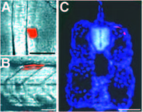
Adaxial cells become the most superficial muscle cells in the somite. (A) Dorsal view of the segmental plate of an approximately 15 h (12 somites) live embryo, after injection of two adaxial cells with lysinated rhodamine dextran. (B) Side view of the same embryo at about 40 h. Both of the injected cells developed into dorsal muscle cells in somite 15 (C) Transverse section of the same embryo, counter stained with Hoechst 33258 to show cell nuclei (blue). The two injected adaxial cells (red) are located superficially. In a series of similar experiments, 72 out of 73 injected adaxial cells became superficial muscle fibers (the single exception was a dorsal cell that may have been adjacent to the neural keel instead of the notochord, and hence misidentified as an adaxial cell at the time of injection). In side views, anterior is to the left; in transverse sections, dorsal is up. Scale bars, 50 μm. |