|
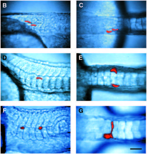
The fate of cells from the notochord domain in flh and ntl mutant embryos. (A) Map of the cells labeled in flh mutant embryos which gave rise to muscle. 10 of these cells fall within the boundary of the notochord domain. In wild-type embryos, 61 of 71 cells labeled within this boundary gave rise to notochord exclusively. The axes of the graph are as in Fig. 3. (B-E,G) flh and (F) ntl mutant embryos during the early pharyngula period, containing clones of cells derived from single labeled cells in the notochord domain of the gastrula. Side (B) and dorsal (C) views of a unilateral clone of muscle cells in a flh embryo. Side (D) and dorsal (E) views of a bilateral muscle clone in a flh embryo. (F) Side view of a clone of axial mesenchymal cells derived from a notochord domain cell in a ntl embryo. (G) Dorsal view of a clone containing a cell that spanned the midline in a flh embryo. Scale bar, 50 μm.
|

