|
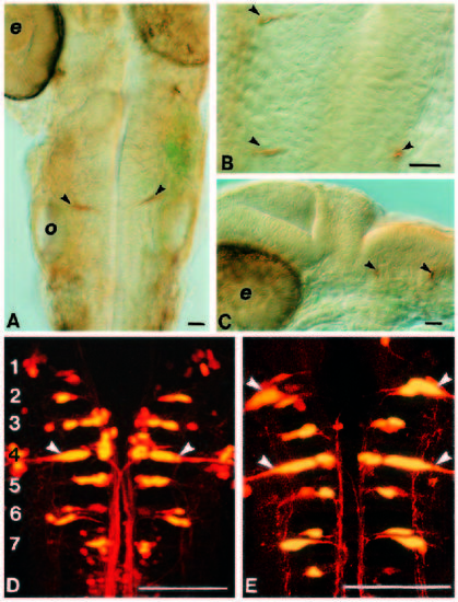
Duplication of the Mauthner neuron occurs in r2 in Hoxa-1- injected embryos. (A-C) 24 hour old embryos stained with the 3A10 antibody. In control embryos (A), a single pair of Mauthner cells is evident in r4 (arrowheads). In injected embryos (B,C), an additional Mauthner cell is seen more rostrally. Anterior is up in A and B and to the left in C. (D,E) Confocal images of hindbrains from larvae that have had lysinated rhodamine dextran crushed onto their spinal cords in order to retrogradely label reticulospinal neurons. (D) Control with the different rhombomeres (numbered) evident from the segmental arrangement of the neurons. The Mauthner neurons is arrowed. Duplicated Mauthner cells are evident in r2 in the injected embryo shown in E. o, otic vesicle; e, eye. Scale bar, 100 μm.
|