
Fig. S6
- ID
- ZDB-FIG-140211-24
- Publication
- Dyer et al., 2014 - A bi-modal function of Wnt signalling directs an FGF activity gradient to spatially regulate neuronal differentiation in the midbrain
- Other Figures
- All Figure Page
- Back to All Figure Page
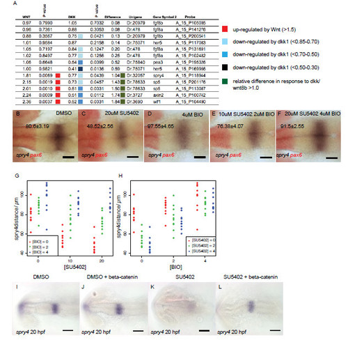
Wnt signalling regulates FGF activity in the midbrain through Sprouty genes. Results from microarray analyses of gene expression showing fold change after upregulation of Wnt8b (column 1) or Dkk1b (column 3) expression (A). P value indicates significance of gene expression changes relative to global changes in gene expression and are colour coded to reflect the fold change. Overall responses to changes in Wnt signalling (genes showing >1.5-fold response to Wnt or Dkk1 upregulation) are represented in column 5 with values >1 representing a consistent response by the gene to changes to Wnt. Dorsal views of spry4 (blue) and pax6 (red) expression in embryos at 20 hpf (values represent distance in μm, ± s.e.m.) treated from 14 hpf with DMSO (B), 20μM SU5402 (C), 4μM BIO (D), 2μM BIO and 10μM SU5402 (E), 4μM BIO and 20μM SU5402 (F). Plots of spry4 expression at 20 hpf after application of varying SU5402 and BIO doses reveals that upregulated Wnt signalling can cause upregulation of spry4 even in the presence of SU5402 (G, H; dots represent individual samples, n=90). Dorsal views of Tg[UAS:HA-bcat]; Tg[hsp70:gal4] embryos processed by in situ hybridisation to examine spry4 expression at 24 hpf after heat-shock at 16.5 hpf and addition of DMSO or 10μM SU5402 (I-L). Scale bars 100μm (I-L), 50μm (A-F). |