
Fig. 3
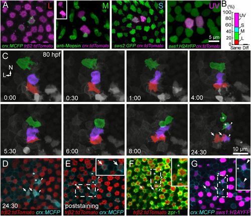
All cone types are generated from symmetric divisions. (A and B) Transient expression of crx:FP produces isolated pairs of labeled cones of the same type. Of 57 pairs, 56 pairs comprised the same cone type (cone pairs observed: 8, L cones; 14, M cones; 9, S cones; 25, UV cones). Only one pair comprised different cone types (L, S). Cone identities were obtained either using cone-specific transgenic lines [Tg(sws2:GFP) Tg(sws1:H2AYFP)] (50) or by M-opsin immunostaining. M-opsin was localized to the outer segments of a pair of crx:tdTomato-expressing cones (Inset). (C) Time lapse of Tg(sws1:H2AYFP, trβ2:tdTomato) fish injected with crx:MCFP at the one-cell stage. Shown here is a region in the dorsal retina where neighboring cell divisions gave rise to pairs of UV, M, and L cones. The precursor and the daughters for each cone type are colorized: UV (violet), M (green), and L (red). Symbols at the last time point in C mark each cone pair that was identified after fixation (D–G). See Movies S4 and S5 for the full time lapse and orthogonal views of the divisions. L, lateral; N, nasal. (D) Single-plane view of the last time point of the live-imaging series in C. (E) One of the cell pairs was trβ2:tdTomato-positive, and, thus, the cells were L cones (arrows). (F) Immunostaining using zpr1 revealed an M-cone pair (asterisks). (G) A UV-cone pair was identified by expression of sws1:H2AYFP (arrowheads). |
| Genes: | |
|---|---|
| Antibody: | |
| Fish: | |
| Anatomical Terms: | |
| Stage Range: | Protruding-mouth to Day 4 |