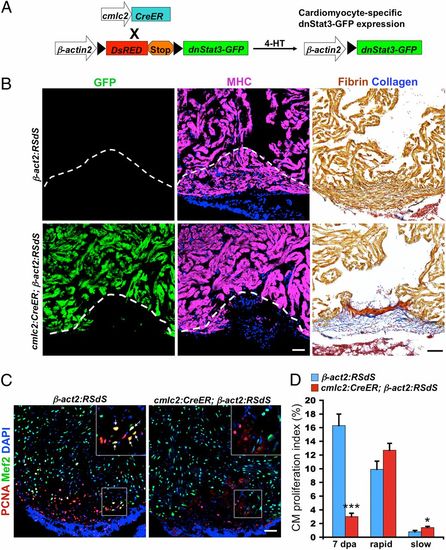
Cardiomyocyte Stat3 activity is essential for heart regeneration. (A) Cartoon representation of transgenes used for inducible expression of a dominant-negative Stat3 (dnStat3-GFP) cassette in cardiomyocytes. (B) cmlc2:CreER;β-act2:RSdS (dnStat3) and β-act2:RSdS (control) clutchmates were administered 4-HT, injured 3 d later, and collected for histological analysis of muscularization and scarring at 30 dpa. Muscle regeneration was blocked, and wounds healed by fibrin retention and scar formation in dnStat3-expressing fish (n = 9). MHC, myosin heavy chain. Dashed lines indicate approximate amputation plane. (C) Confocal images of sections from 7dpa ventricles, stained with antibodies against PCNA (a proliferation marker) and Mef2 (a cardiomyocyte marker). (Insets) Higher-magnification images of the squares; arrows, proliferating cardiomyocytes. (D) Quantification of cardiomyocyte (CM) proliferation during regeneration (7 dpa) or after 10 d of rapid or slow (normal) growth conditions. Ventricular resection in adults triggered similar levels of cardiomyocyte proliferation as rapid growth conditions in juveniles/young adults. However, inhibitory effects of dnStat3 expression were apparent only during injury-induced regeneration. Data are mean ± SEM n = 6 (regeneration) or 12 (growth), *P < 0.05, ***P < 0.001. Student t test (unpaired, two-tailed). (Scale bars, 50 μm.)
|
