
Fig. S1
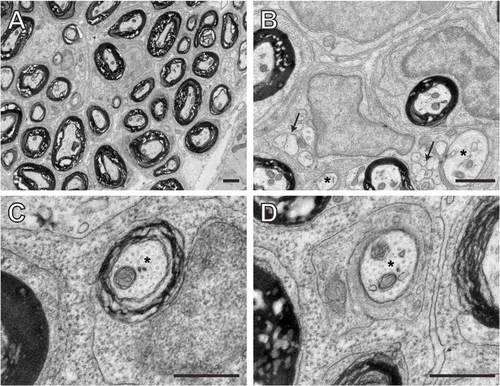
Schwann cells at different developmental stages in the posterior lateral line nerve at 50 dpf. (A-D) TEM images of transverse sections through adult wild-type zebrafish at 50 dpf showing the ultrastructure of the PLL nerve. (A) Low magnification view of the PLLn showing many myelinated axons. (B-D) Higher magnification views of the PLLn show Schwann cells at multiple stages of development. The arrows in B mark groups of unsorted axons that are ensheathed by immature Schwann cells, and the asterisks mark sorted axons. Multiple myelinated axons are also visible in the same field. (C) A myelinating Schwann cell has elaborated several wraps around an axon (marked with asterisk) although the level of myelination is similar to that observed at earlier stages (compare with Fig. 1). (D) A Schwann cell has established a 1:1 relationship with an axon (marked with asterisk), but has not yet initiated myelination. Scale bars: 1 μM in A,B; 0.5 μM in C,D. |