Fig. 1
- ID
- ZDB-FIG-130815-5
- Publication
- Gupta et al., 2013 - Loss of Catalytically Inactive Lipid Phosphatase Myotubularin-related Protein 12 Impairs Myotubularin Stability and Promotes Centronuclear Myopathy in Zebrafish
- Other Figures
- All Figure Page
- Back to All Figure Page
|
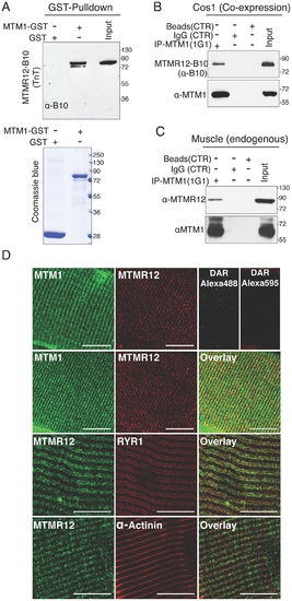
Protein-protein interactions between myotubularin and MTMR12 proteins. (A) GST pull down of MTM1-GST recombinant protein with MTMR12 showed a direct interaction between the two proteins (above). Coomassie blue stained gel (below) showing GST and MTM1-GST protein used in the pull down (B) Co-IP experiments from Cos-1 transfected cells with MTM1 and MTMR12-B10 constructs showing the interaction of the two proteins in the cellular context in-vivo (C) MTMR12 co-immunoprecipitates with myotubularin (using 1G1 monoclonal antibody) in mouse muscle lysates revealed with anti-MTMR12 polyclonal antibody (upper panel) and with the 2827 polyclonal anti-myotubularin (bottom panel). (D) Confocal microscopic immunofluorescence studies of longitudinal frozen sections of skeletal muscle from mouse tibialis anterior muscle with sarcomeric markers. Individual immunostaining with 2827 anti-myotubularin or anti-MTMR12 showed similar striated localization in skeletal muscle (top panel). Double-immunostaining with both proteins showed a co-localization of myotubularin and MTMR12 to Triads and partial co-localization with the ryanodine receptor, RyR1, a sarcoplasmic reticulum marker, but not with α-actinin, a Z-line marker. Scale bar = 10 μm. |

