
Fig. 4
- ID
- ZDB-FIG-130813-8
- Publication
- Eames et al., 2013 - FishFace: interactive atlas of zebrafish craniofacial development at cellular resolution
- Other Figures
- All Figure Page
- Back to All Figure Page
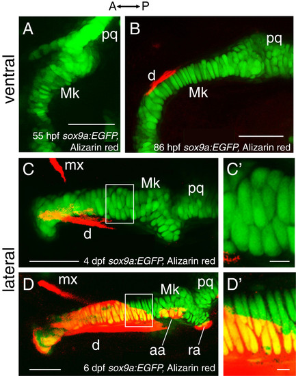
Chondrocyte intercalation and proliferation seem to drive cartilage growth in late embryonic and early larval stages. Confocal images in ventral view of Alizarin red-stained sox9a:EGFP mandibles suggest that early phases of Meckel’s cartilage morphogenesis may be achieved by chondrocyte intercalation. By 55 hpf (A), chondrocytes in Meckel’s cartilage do not appear in ordered rows, as generally two to three cells span the mediolateral width of the element. By 86 hpf (B), Meckel’s cartilage has grown in length and is relatively thinner from that seen at 55 hpf, and many of its chondrocytes now span the mediolateral width in a cellular stack. Confocal images in lateral view of Alizarin red-stained sox9a:EGFP mandibles suggest that later phases of Meckel’s cartilage morphogenesis may be achieved by chondrocyte proliferation. Chondrocytes of Meckel’s cartilage are separated clearly from each other by 4 dpf (C). By 6 dpf, proliferation of chondrocytes about mid-way along the anterior-posterior length of Meckel’s cartilage (D) is suggested by the presence of chondrocyte doublets. These doublets are separated from each other by a layer of presumed extracellular matrix that appears much thinner than that observed between the doublets. C′ and D′ show high magnification views of the boxed regions in C and D, respectively. Abbreviations: A = anterior; aa = anguloarticular; d = dentary; dpf = days post-fertilization; hpf = hours post-fertilization; Mk = Meckel’s cartilage; mx = maxilla; P = posterior; pq = palatoquadrate; ra = retroarticular. Scale bars: A-D = 50 μm; C′,D′ = 5 μm. |