
Fig. 1
- ID
- ZDB-FIG-130801-35
- Publication
- Das et al., 2013 - Participation of PI3-kinase/Akt signalling in insulin stimulation of p34cdc2 activation in zebrafish oocyte: Phosphodiesterase 3 as a potential downstream target
- Other Figures
- All Figure Page
- Back to All Figure Page
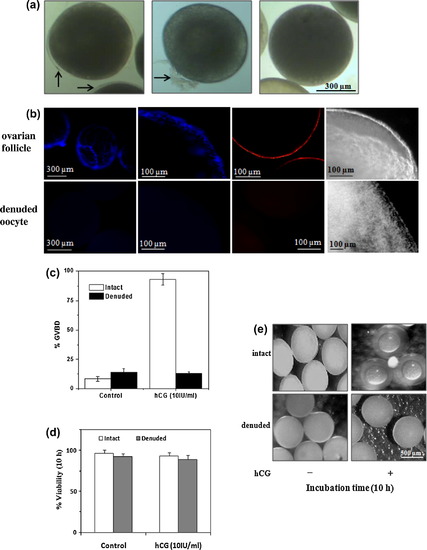
Representative micrographs of fully grown zebrafish oocytes (mean diameter <650 μm) at various steps during removal of follicular wall components due to collagenase treatment (a). Arrows indicate follicle layer. Comparison of surface images of fully grown ovarian follicles (upper panel) with denuded oocytes (lower panel) either stained with DAPI or PI under an upright-fluorescent microscope (b). Corresponding phase contrast images at higher magnification are also shown (extreme right). DAPI or PI positive cells reveal the presence of follicle layer. The intact follicles and denuded oocytes were incubated in vitro with hCG (10 IU/ml of culture media) for 10 h and scored for GVBD (c) and percent viability (d). Representative micrographs of the cultured follicles and collagenase-treated oocytes after 10 h of incubation recorded in reflected light against the dark background under stereo zoom microscope (e). |
Reprinted from Molecular and Cellular Endocrinology, 374(1-2), Das, D., Khan, P.P., and Maitra, S., Participation of PI3-kinase/Akt signalling in insulin stimulation of p34cdc2 activation in zebrafish oocyte: Phosphodiesterase 3 as a potential downstream target, 46-55, Copyright (2013) with permission from Elsevier. Full text @ Mol. Cell. Endocrinol.