
Fig. 3
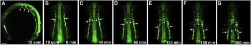
The Migration of Trunk Angioblasts in Live etv2:GFP-Transgenic Embryos(A–G) GFP expression is observed in the medial and lateral endothelial progenitor cells at the 10- to 16-somite stages. In laterally (A) and dorsally mounted embryos (B–G), medial angioblasts (white arrows) migrate toward the midline (white arrowheads) intersomitically. (B–G) Selected frames from a time-lapse movie showing migration of the medial angioblasts; imaging was started at the 10-somite stage. The anterior angioblasts migrate first, followed by the more posterior endothelial progenitor cells. (F and G) Faint GFP expression in the lateral angioblasts (red arrowheads) becomes apparent; these cells migrate to the midline similar to the medial angioblasts. In (A) anterior is to the left; in (B)–(G) anterior is up. Scale bar, 100 μm. See also Figure S2 and Movies S1, S2, S3, and S4. |
Reprinted from Developmental Cell, 25(2), Kohli, V., Schumacher, J.A., Desai, S.P., Rehn, K., and Sumanas, S., Arterial and venous progenitors of the major axial vessels originate at distinct locations, 196-206, Copyright (2013) with permission from Elsevier. Full text @ Dev. Cell