
Fig. 4
- ID
- ZDB-FIG-130516-41
- Publication
- Lévesque et al., 2013 - Inflammation drives wound hyperpigmentation by recruiting pigment cells to sites of tissue damage
- Other Figures
- All Figure Page
- Back to All Figure Page
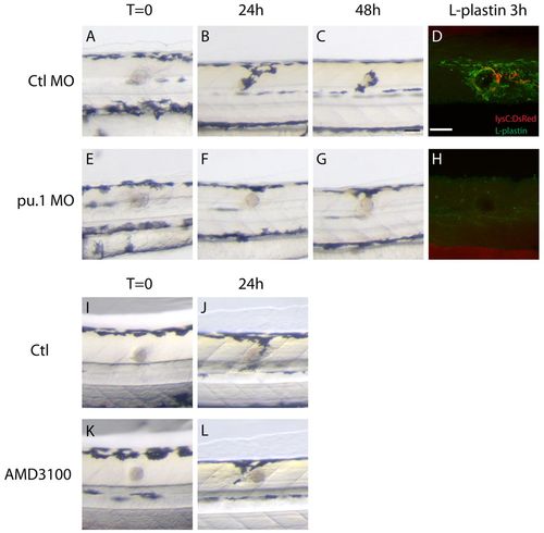
Wound recruitment of melanocytes requires innate immune cells. (A-H) Time-course of melanocyte recruitment to the bead in control and pu.1 MO-injected fish. (A-C) As shown in Fig. 1, there is a time-dependent migration of melanocytes to the wound over the first 48 hours following bead implantation in control fish. (E-G) Depletion of innate immune cells in pu.1 MO-injected fish delays and reduces the recruitment of melanocytes to the wound. (D) Immunostaining of innate immune cells with anti-L-plastin antibody shows the presence of leukocytes around the wound in control fish; neutrophils (lysC:DsRed) are red and macrophages (L-plastin-positive cells) are green. (H) No leukocytes are evident in pu.1 MO-injected fish. (I-L) Compared with control fish (I,J), AMD3100-treated fish do not show a reduction in wound hyperpigmentation at 24 hpi (K,L). Scale bar: 50 μm (A-L). |