
FIGURE
Fig. S2
- ID
- ZDB-FIG-130502-9
- Publication
- Muto et al., 2013 - Real-Time Visualization of Neuronal Activity during Perception
- Other Figures
- All Figure Page
- Back to All Figure Page
Fig. S2
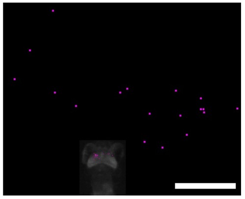
Calcium Imaging with a Free-Swimming Zebrafish Larvae during Perception of a Paramecium, Related to Figure 4 Positions of Ca2+ signals (small dots in the optic tectum of the larvae) and a swimming paramecium (large dots in the surrounding area) just before the start of approach swimming. Results were obtained from 18 recordings. Scale bar: 1000 μm. |
Expression Data
Expression Detail
Antibody Labeling
Phenotype Data
Phenotype Detail
Acknowledgments
This image is the copyrighted work of the attributed author or publisher, and
ZFIN has permission only to display this image to its users.
Additional permissions should be obtained from the applicable author or publisher of the image.
Full text @ Curr. Biol.