|
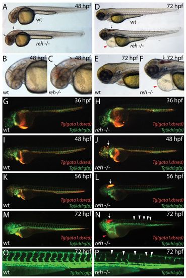
reddishs587 mutants exhibit cranial hemorrhage, degenerating vessels and pericardial edema. (A-F) Bright-field micrographs of wild-type (wt) and rehs587 mutant (reh-/-) embryos at (A-C) 48 hpf and (D-F) 72 hpf (A,C,D,F). Black arrows indicate cranial hemorrhage in reh mutant. Red arrowheads indicate pericardial edema. (G-N) Fluorescence micrographs of (G,I,K,M) wild-type (wt) and (H,J,L,N) rehs587 mutants (reh-/-) in Tg(gata1:dsRed);Tg(kdrl:GFP) background at (G,H) 36, (I,J) 48, (K,L) 56 and (M,N) 72 hpf. Cranial hemorrhage as detected by extravasation of Tg(gata1:dsRed) labeled blood in the head (white arrows) occurs in rehs587 mutants as early as (J) 48 hpf and increases during development (L,N). reh-/- intersegmental vessels appear to degenerate by 72 hpf (N, white arrowheads). (O,P) Tg(kdrl:GFP) (O) wild-type (wt) and (P) reh-/- intersegmental vessels confirm this degeneration in reh-/- mutants at 72 hpf (white arrowheads). Red arrowhead in N indicates pericardial edema.
|

