
Fig. 3
- ID
- ZDB-FIG-130403-5
- Publication
- Tan et al., 2013 - Spatiotemporal expression of the dermatopontin gene in zebrafish Danio rerio
- Other Figures
- All Figure Page
- Back to All Figure Page
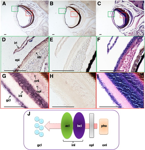
In situ hybridization of dermatopontin (DPT) mRNA in adult zebrafish eye. In this and other figures, unless otherwise mentioned, sections were transversely cut with the dorsal on top. Sections (8 μm) were hybridized with the DPT probes. Scale bar = 100 μm. (A) Hybridization of the antisense DPT probe. (B) Hybridization of sense DPT probe. (C) Hematoxylin and eosin staining. Higher magnification images of the green box and red box in (A, B, and C) are shown in (D, G), (E, H), and (F, I), respectively. (J) A schematic diagram showing the process of optic nerve signal transmission. Abbreviations: iri, iris; epi, epithelium; str, stroma; gcl, ganglion cell layer; inl, inner nuclear layer; opl, outer plexiform layer; onl, outer nuclear layer; acl, amacrine cell layer; bcl, bipolar cell layer; pho, photoreceptors. |
| Gene: | |
|---|---|
| Fish: | |
| Anatomical Terms: | |
| Stage: | Adult |
Reprinted from Gene, 516(2), Tan, Y., Iimura, K., Sato, T., Ura, K., and Takagi, Y., Spatiotemporal expression of the dermatopontin gene in zebrafish Danio rerio, 277-284, Copyright (2013) with permission from Elsevier. Full text @ Gene