
Fig. 4
- ID
- ZDB-FIG-130228-31
- Publication
- Mikelsaar et al., 2012 - Epitope of titin a-band-specific monoclonal antibody Tit1 5 H1.1 is highly conserved in several Fn3 domains of the titin molecule. Centriole staining in human, mouse and zebrafish cells
- Other Figures
- All Figure Page
- Back to All Figure Page
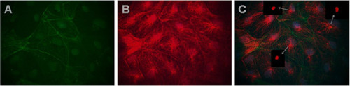
Immunofluorescence co-localization of MAb Tit1 5 H1.1 target antigen (titin) with F-actin in different cells of zebrafish primary culture of the testes. Cells were fixed on 3rd day of cultivation with 4% PFA, permeabilized, and double-labelled (C) with Mab Tit1 5 H1.1 for titin (red - Alexa 594) and with Phalloidin for F-actin (green – Alexa 488). Cell nuclei were stained blue with DAPI (obj. 100x). Arrows show centriole staining.In Photoshop image processing either red colour (A) or green colour (B) was removed and one can see independent staining both of F-actin (A) and titin (B). Note a very extensive fibrous centriole-orientated staining of titin. |
| Gene: | |
|---|---|
| Antibody: | |
| Fish: | |
| Anatomical Terms: | |
| Stage: | Adult |