
Fig. 4
- ID
- ZDB-FIG-130211-5
- Publication
- Nair et al., 2013 - In vitro oocyte culture-based manipulation of zebrafish maternal genes
- Other Figures
- All Figure Page
- Back to All Figure Page
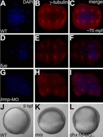
Stage IV oocyte injections of Lrmp and Dhx16 morpholinos phenocopy fue and mis maternal-effect phenotypes, respectively. A–I: Animal views of immunolabeled 70 minutes postfertilization (mpf) blastodiscs stained for DAPI (A,D,G) immunolabeled to detect γ-tubulin (B,E,H) and panel merges (C,F,I). A–C: In wild-type embryos, each nucleus associates with centrosomal γ-tubulin staining. D–F: In maternal-effect fue mutants, nuclei fail to divide, resulting in two to three patches of nuclei stainings corresponding to unfused parental pronuclei and the polar body for meiosis II (D,F), which fail to associate with γ-tubulin (E,F). G–I: In Lrmp morphants where maternal Lrmp function was inhibited, the nuclei similarly fail to divide (G) and also fail to associate with γ-tubulin (H,I). J–L: Lateral views of live embryos at 9 hours postfertilization (hpf). J,K: Wild-type embryos at 9 hpf where axis formation is evident (anterior to the top and dorsal to the right) (J), while in maternal-effect mis mutants, epiboly fails to occur (K). L: In Dhx16 morphants where maternal function was inhibited, epiboly similarly arrests, resulting in gastrulation failure. |