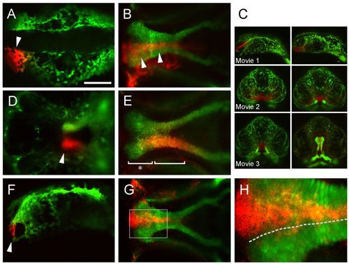
Palate morphogenesis in zebrafish. (A,B) The anterior cells at 14-somites (arrowhead, A) tracked to the median ethmoid plate at 4.5 dpf (arrowheads, B). (C) Screenshots of time-lapse movies capturing this process. (D,E) Unilateral labeling of the entire maxillary prominence at 55 hpf (arrowhead, D) revealed that the mature lateral ethmoid plate and trabeculae are formed both by uniform expansion (bracket, E) as well as increased proliferation at the leading edge (asterisk) (see also Fig. 2C, supplementary material Movie 4). (F,G) Photoconversion of FNP cells at 20 somites (F arrowhead) labeled only the median ethmoid at 4.5 dpf (G), also captured in supplementary material Movies 1-3. (H) Enlargement of inset in G. Inspection of the ethmoid plate revealed that labeled CNCCs could be traced along the seam (broken line) between the median and lateral ethmoid plate; lateral to this line, chondrocytes are columnar in appearance while medially they are cuboidal. A, dorsal view; F, lateral view; B,D,E,G,H, ventral views. Scale bar: 50 μm.
|
