
Fig. 1
- ID
- ZDB-FIG-130115-19
- Publication
- Watanabe et al., 2012 - In vivo assessment of the permeability of the blood--brain barrier and blood-retinal barrier to fluorescent indoline derivatives in zebrafish
- Other Figures
- All Figure Page
- Back to All Figure Page
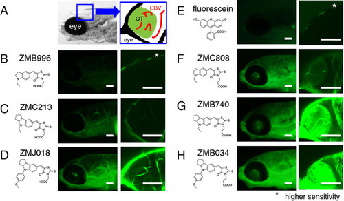
In vivo assessment of the permeability of the BBB to fluorescent compounds. Zebrafish larvae (casper line) at 7–8 dpf were immersed in egg water containing 1 μM of an ID or fluorescein for 1 h. In vivo fluorescence imaging of the zebrafish brain was performed using fluorescence microscopes. A: Schematic diagram showing the region observed using the fluorescence microscopes. B-H: In vivo fluorescence imaging of zebrafish larvae stained with ID possessing a rhodanine ring with an acetic acid group (ZMB996, ZMC213, and ZMJ018, B, C and D, respectively), with fluorescein (E), and with ID possessing a rhodanine ring with a propanoic acid group (ZMC808, ZMB740, and ZMB034, F, G and H, respectively). The OT was clearly visualized in zebrafish stained with IDs possessing a rhodanine moiety with a propanoic acid group. Scale bar: 100 μm. OT, optic tectum; CBV, cerebral blood vessel. |