
Fig. 4
- ID
- ZDB-FIG-121220-13
- Publication
- Walogorsky et al., 2012 - Zebrafish model for congenital myasthenic syndrome reveals mechanisms causal to developmental recovery
- Other Figures
- All Figure Page
- Back to All Figure Page
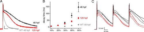
The effects of accelerated synaptic current decay on membrane repolarization. (A) Muscle cell voltage responses to the single firing of a motor neuron. Between 12 and 20 individual muscle responses were averaged to generate the average waveforms corresponding to 48-hpf wild-type (WT; gray), 48-hpf twi+/− (black), and 120-hpf twi+/− (red) fish. The scale bar represents 10 mV and 10 ms. (B) Comparison of the repolarization time course among 48-hpf wild-type (gray), 48-hpf twi+/− (black), and 120-hpf (red) twi+/− animals. (C) Sample traces for the first three sequential muscle responses to motor neuron firing at 20 Hz. Repolarization was incomplete for both 48-hpf (black traces) and 120-hpf (red traces) twi+/− fish. However, the take-off level for responses 2 and 3 was much more positive at 48 hpf. |
| Fish: | |
|---|---|
| Observed In: | |
| Stage: | Long-pec |