
Fig. 4
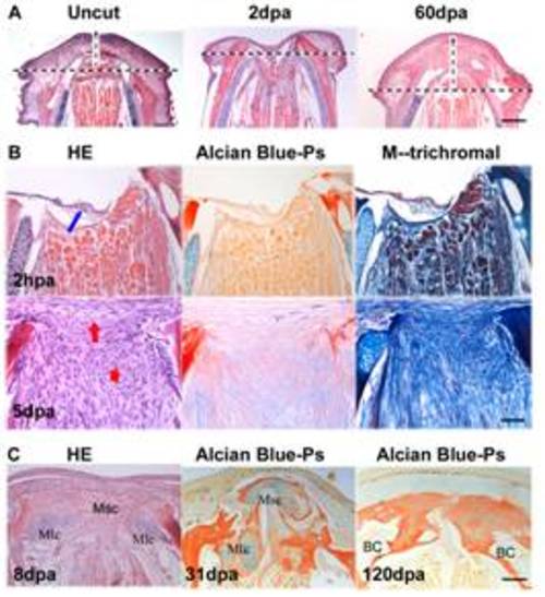
Histological observation of blastema formation and reformation. Figure A shows that regeneration of the mandible retains the original mandibular arch. The cut arch (arrow) at 2 dpa was restored to the original shape (uncut) at 60 dpa. HE staining. Scale bars, 250 μm. Figure B shows blastemal structure and extracellular matrix composition. At 2 hpa, wound epidermis reconstitution started (blue arrow), and blastema was not yet formed between wound epidermis and the injured muscle. At 5 dpa, the blastemal ECM began to reorganize toward hypodermis (vertical red arrow) and the chondrogenic center (horizon red arrow). In comparison with HE stains, Alcian blue-Ponceau S staining shows hyaluronic acid and hyaline cartilage as blue, collagen and mineralized bone matrix as red; In a modification of Masson′ trichromal staining, green or blue staining is collagen; brown staining is elastic fibers; red is cytoplasm, muscle, nerve sheathe, fibronectin and erythrocytes). Scale bars, 50 μm. Figure C shows two types of chondrogenic ossification. After blastema formation (8 dpa), chondrogenic blastema was composed of three chondrogenic ossification centers: two Meckel-lateral centers (Mlc) and one median symphysis center (Msc). In the Meckel-lateral centers, perichondral ossification was more like atypical or incomplete endochondral ossification. The surrounding matrix became calcified while the central portion of ossification center developed to bone cysts (BC), a similar structure like bone marrow cavity filled with little connective tissues (120 dpa). The median symphysis center, however, adopted perichondral ossification. Scale bars, 100 μm. |