
Fig. 2
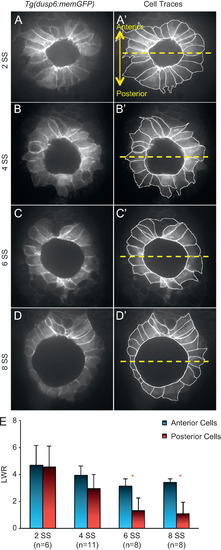
Ciliated KV cells undergo region-specific cell shape changes during morphogenesis. (A–D) Optical cross-sections through the middle focal plane of KV cells expressing membrane-localized GFP in wild-type Tg(dusp6:memGFP) embryos at different stages of development. (A′–D′) KV cell boundaries were outlined to highlight cell shapes. The dashed yellow line bisects KV into anterior and posterior regions. All images are oriented with anterior at the top and posterior at the bottom. (E) Length-to-width ratio (LWR) of anterior and posterior cells at different stages of development. Error bars=one standard deviation. n=number of embryos analyzed. * Significant difference between anterior and posterior (p<0.01). |
Reprinted from Developmental Biology, 370(1), Wang, G., Manning, M.L., and Amack, J.D., Regional Cell Shape Changes Control Form and Function of Kupffer's Vesicle in the Zebrafish Embryo, 52-62, Copyright (2012) with permission from Elsevier. Full text @ Dev. Biol.