
FIGURE
Fig. 4
- ID
- ZDB-FIG-121004-13
- Publication
- Kaufmann et al., 2012 - Multilayer mounting enables long-term imaging of zebrafish development in a light sheet microscope
- Other Figures
- All Figure Page
- Back to All Figure Page
Fig. 4
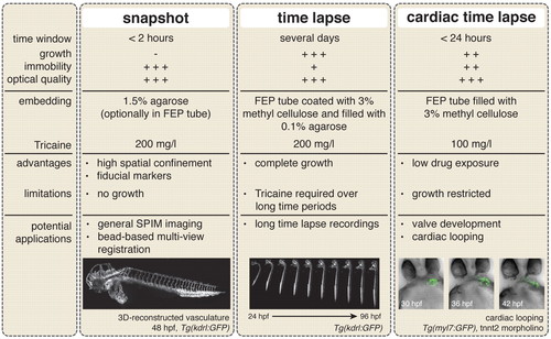
Recommended mounting protocols for light sheet microscopy. Three different experimental settings and their potential applications. The image of the 3D-reconstructed vasculature is a maximum projection (supplementary material Movie 2). Images of the developing zebrafish embryo in 0.1% agarose and the developing heart in 3.0% methylcellulose are taken from supplementary material Movies 1 and 3, respectively. |
Expression Data
Expression Detail
Antibody Labeling
Phenotype Data
Phenotype Detail
Acknowledgments
This image is the copyrighted work of the attributed author or publisher, and
ZFIN has permission only to display this image to its users.
Additional permissions should be obtained from the applicable author or publisher of the image.
Full text @ Development