
Fig. 6
- ID
- ZDB-FIG-120907-29
- Publication
- Salbreux et al., 2012 - Coupling mechanical deformations and planar cell polarity to create regular patterns in the zebrafish retina
- Other Figures
- All Figure Page
- Back to All Figure Page
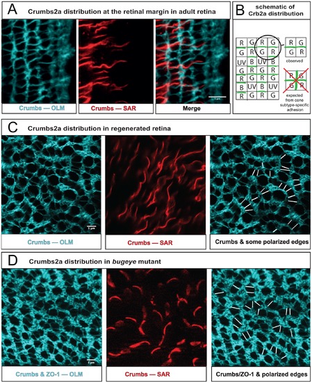
Planar cell polarity in intact and regenerated retina. A) Crb2a protein localized by immunocytochemistry in a flat-mount preparation at the margin of the adult retina (germinal zone to the right). The focal plane is at the level of the zonula adherens/OLM (left panel, projection of 12 confocal z slices, cyan); at the level of the inner segments/SAR of cone photoreceptors (middle panel, single confocal z slice, red); an overlay of both panels (right). See also Fig. S4J–L. B) Schematic illustrating Crb2a distribution along cone-cone interfaces within a column at the level of the SAR. Note that Crb2a does not localize to the orthogonal interfaces between adjacent cones across columns, as would be expected if Crb2a mediated unpolarized, but spectral-subtype-dependent, interactions between cones. C) At the level of the OLM, cone photoreceptors (large profiles) in regenerated retina are not organized in a rectangular lattice, but aggregate into short chains one cell wide, indicating polarized interactions (left panel). The planar polarized interfaces are verified by Crb2a localization in the SAR of these cones (middle panel). By tracing the Crb2a signal through successive focal planes in the Z-stack some planar polarized SAR interfaces between cone inner segments were associated with the corresponding cone-cone interfaces at the OLM, as indicated by white line segments (right panel). D) At the level of the OLM, cone profiles in the adult bugeye mutant are organized similarly to the regenerated retina, here visualized with a cocktail of antibodies against ZO-1 and Crb2a (left panel). Again similar to the regenerated retina, Crb2a localizes to planar polarized SAR interfaces between cone inner segments (middle panel), and some of these were traced to the cone-cone interfaces at the OLM (right panel). |