
Fig. S1
- ID
- ZDB-FIG-120814-3
- Publication
- Zhou et al., 2012 - ALDH2 Mediates 5-Nitrofuran Activity in Multiple Species
- Other Figures
- All Figure Page
- Back to All Figure Page
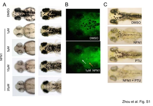
Melanocyte sensitivity to NFN1 treatment. A. Example images of zebrafish following increasing concentrations of NFN1 (left panel), and transfer to fresh E3 embryo medium (washout) for three days. B. Melanocytes are visible in transgenic zebrafish embryos expressing melanocyte specific tryp1-GFP both by the black pigment and GFP poistive cytoplasm. In NFN1 treated embryos most melanocytes are no longer visible, and black detritus is not associated with GFP positive cytoplasm (white arrow), although a few melanocytes survive (orange arrow). C. PTU does not rescue NFN1 activity in melanocytes. PTU inhibits the activity of tyrosinase, the rate-limiting enzyme required for pigment synthesis. Two dpf zebrafish embryos were pre-treated with 30mg/l PTU for 6 hours, and then treated with 0.5 μM NFN1 or DMSO as a control. |