FIGURE

Fig. 2

ID
ZDB-FIG-120810-33
Publication
Johnson et al., 2012 - Scube activity is necessary for Hedgehog signal transduction in vivo
Other Figures
All Figure Page
Back to All Figure Page
Fig. 2

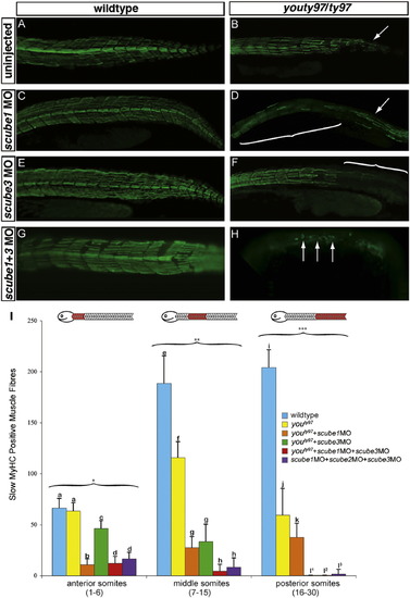
scube1 and scube3 morpholinos enhance the phenotype of youty97 mutant embryos. The wildtype pattern of slow MyHC (A) is disrupted in the posterior tail region of youty97 homozygotes (B) (arrow). The pattern of slow MyHC is not visibly affected when scube1MO (C) scube3MO (E) or both scube1MO+scube3MO (G) are injected into wildtype embryos. (D) scube1MO injected into youty97 homozygotes enhanced the youty97 phenotype. Slow MyHC expression is disrupted after youty97 homozygotes in this region (compare B to D). Bracket highlights the region in youty97+scube1MO embryos lacking slow MyHC fibers. An arrow indicates the somitic region where slow MyHC expression resembles youty97 mutants. (F) scube3MO injected into youty97 homozygotes also enhanced the youty97 mutant phenotype. Slow MyHC is disrupted after <8–10 somites, all remaining somites completely lack slow MyHC expression. Bracket highlights the somitic region in youty97+scube3MO embryos devoid of slow MyHC staining. (H) Injection of scube1MO+scube3MO together into youty97 mutant embryos essentially eliminates slow MyHC staining. Arrows highlight the few remaining slow MyHC positive fibers remaining in youty97+scube1MO+scube3MO embryos (H). Scube triple MO injection into wildtype embryos faithfully reproduces the phenotype seen in youty97+scube1MO+scube3MO (H and data not shown). All embryos<30 hpf. F59 monoclonal antibody labels slow MyHC positive muscle fibers, Alexa 488-conjugated donkey anti-mouse secondary. (I) Slow MyHC positive muscle fibers were counted in anterior (1–6 somites), middle (7–15 somites) and posterior (16–30) somites of wildtype, youty97 homozygous mutants, youty97+scube1MO embryos, youty97+scube3MO embryos, youty97+scube1MO+ scube3MO embryos, and scube triple MO embryos (scube1MO+scube2MO+scube3MO). Statistical analysis demonstrated the phenotypic enhancement of the youty97 mutant phenotype seen following MO injection is significant. The dramatic loss of slow MyHC positive muscle fibers seen following scube1MO+scube3MO injection into youty97 homozygotes was equivalent to the loss of slow MyHC positive muscle fibers seen following scube triple MO injection into wildtype embryos (l1-3). *ANOVA F(5,137)=200.11, p<0.001. **ANOVA F(5,137)=266.10, p<0.001. ***ANOVA F(5,137)=391.35, p<0.001. Within each somite region, bars with different letters are statistically significant from one another.

Expression Data

Expression Detail
Antibody Labeling
Phenotype Data
Fish:
Knockdown Reagents:
Observed In:
Stage: Prim-5

Phenotype Detail
Acknowledgments
This image is the copyrighted work of the attributed author or publisher, and ZFIN has permission only to display this image to its users. Additional permissions should be obtained from the applicable author or publisher of the image.

Reprinted from Developmental Biology, 368(2), Johnson, J.L., Hall, T.E., Dyson, J.M., Sonntag, C., Ayers, K., Berger, S., Gautier, P., Mitchell, C., Hollway, G.E., and Currie, P.D., Scube activity is necessary for Hedgehog signal transduction in vivo, 193-202, Copyright (2012) with permission from Elsevier. Full text @ Dev. Biol.