
Fig. 3
- ID
- ZDB-FIG-120810-11
- Publication
- Lee et al., 2012 - Development of a transient expression assay for detecting environmental oestrogens in zebrafish and medaka embryos
- Other Figures
- All Figure Page
- Back to All Figure Page
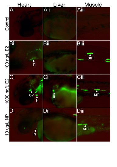
Comparison of detectable response concentrations for different oestrogen compounds in the zebrafish embryo transient assay. Injected embryos were exposed for up to 72 h post-fertilisation to 17β-oestradiol (E2) and nonylphenol (NP) and fluorescence detected using fluorescence microscopy (Leica DMI 4000 B). No GFP expression was observed in controls (A1-A3), but low level GFP expression occurred in the liver for exposure to 1000 ng E2/L (C2) and high level GFP expression was detected in the somite muscle and heart at exposure concentrations of 100 and 1000 ng E2/L. Weak GFP expression was also detected in the heart and somite muscle of embryos for exposure to 10 μg/L NP (D1-D3). Embryos exposed to the higher concentrations of NP (100 μg/L) died. This experiment was run in duplicate and was repeated at least seven times. |