
Fig. 3
- ID
- ZDB-FIG-120803-4
- Publication
- Talbot et al., 2012 - fras1 shapes endodermal pouch 1 and stabilizes zebrafish pharyngeal skeletal development
- Other Figures
- All Figure Page
- Back to All Figure Page
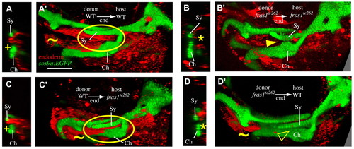
Endodermal fras1 sculpts epithelia and skeleton. Confocal transverse sections taken at a section level midway through Sy length at 7 dpf. Medial to right, dorsal up. (A) In mosaic fish in which WT endoderm has been transplanted into WT hosts (WT→WT mosaics), late-p1 appears normal (+) and is labeled as endoderm. (B) In fras1te262→fras1te262 mosaic fish, late-p1 does not form, and anterior endoderm remains medial to cartilages (asterisk). (C) WT endoderm transplanted into fras1te262 mutants rescues the late-p1 defects of these hosts. (D) Half of the fras1te262→WT mosaics examined exhibited defective endoderm medial to Sy and Ch. (A′-D′) Rendered confocal stacks show endoderm and skeletal morphology; oriented anterior to the left, dorsal up. Image rendering makes both tissues opaque, allowing visualization of late-p1 covering separated Sy and Ch cartilages (circled). In WT→WT (A′) and WT→fras1te262 (C′) mosaics, late-p1 covers part of Ch. However, in fras1te262→fras1te262 mosaics, fused cartilages cover the medial endoderm (arrowhead). Half of the fras1te262→WT mosaics show late-p1 defects, but cartilage defects are more subtle than in non-mosaic fras1te262 mutants, perhaps owing to the presence of late-p1 in the first pharyngeal arch (tilde). Scale bar: 100 μm. |